In situ microscopy is an investigatory technique that allows researchers to observe how a sample reacts to stimuli under an electron microscope. Before in situ microscopy, researchers were not able to witness real-time effects; they were blind to reactions and processes as they happened and could only see end results.
 The real-time observational capabilities of in situ microscopy have supported revolutionary discoveries in numerous areas of study. In situ microscopy is used to observe reactions in a variety of research areas including life sciences, catalysis, materials characterization, batteries, and much more.
In situ microscopy is complex and involves numerous processes and tools, as well as advanced scientific study and training for microscope operations. This glossary can help clarify in situ microscopy terms for those new to the research technique.
Electron Microscope (EM)
An electron microscope can deliver magnifications hundreds to thousands of times larger than light microscopes by imaging samples with a beam of electrons. They are widely used by researchers to study fine detail in a variety of samples and Ems generally require trained personnel to operate.
Transmission Electron Microscope (TEM)
A transmission electron microscope (TEM) is a type of electron microscope that employs a high voltage electron beam, which is emitted by a cathode and formed by magnetic lenses. The electron beam is simultaneously transmitted through the specimen, creating an image that can illuminate the size and structure of the sample. Magnetic lenses then magnify the image and capture a digital image which is two-dimensional, black and white, and can be displayed in real-time on a computer monitor.
Scanning Electron Microscope (SEM)
A scanning electron microscope (SEM) directs an electron beam at a sample and captures an image based on elastically or inelastically scattered electrons emitted via excitation from the surface of the sample. In this type of microscope, the electron beam scans across the sample in a raster pattern, and an image is created from the signals detected during the scanning process.
Scanning Transmission Electron Microscope (STEM)
A scanning transmission electron microscope (STEM) combines TEM and SEM techniques; like a TEM, an electron beam is projected through a sample. But, like a SEM, the electron beam moves through incremental areas, creating an image by combining data from these small sections. An image is then compiled and displayed in real-time on a monitor.
Energy-Dispersive X-Ray Spectroscopy (EDS)
EDS is a technique used for chemical characterization of a sample. Since each element emits characteristic x-rays when exposed to a high energy electron beam, this technique is able to determine the composition of samples. EDS can be used in electron microscopes, SEM, and STEM.
EDS Detector
This device is used for EDS chemical characterization. It converts x-ray energy into voltage signals, which are then sent to a pulse processor. The pulse processor measures the signals and passes them on for data display and analysis.
Environmental TEM (ETEM)
ETEM describes the practices and products used to introduce a gaseous environment into a TEM. ETEM was first introduced as a microscope equipped with gas nozzles that would allow researchers to pump gas into the sample. Now, ETEM is mostly used as specialized in situ microscopy holders that allow for the study of native gas environments.
TEM Holder
TEM holders are devices used within an electron microscope to house and support a sample. In situ TEM holders can alter the sample’s environment – by heat, liquid, or gas – to allow for native sample study.
Sample Support
Traditionally, the very small sample size needed for TEM analysis requires samples be supported by some structure so that they do not fall into the vacuum of the TEM. Sample supports come in a wide variety of shapes and sizes, and use many different materials as the support structure.
Microelectromechanical Systems (MEMS)
MEMS are a technology defined as miniaturized mechanical and electro-mechanical elements made with microfabrication. MEMS contain microsensors and microactuators, which are transducers that convert energy from one form into another. Typically, this involves converting a mechanical signal into an electrical signal. MEMS are used in electron microscopes to change the sample environment of experiments.
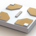
E-chips
E-chips are the proprietary name for Protochips MEMS-based sample supports and are used to support and confine in situ samples. E-chips are used for in situ TEM holders, including Protochips’ Atmosphere, Fusion, and Poseidon Select products.
Membrane
E-chips and other MEMS devices used in closed cells require very thin sample supports that remain transparent to the electron beam and can confine liquids and gasses. A common membrane support would be amorphous Silicon Nitride (SiN) about 30 nm thick.
High-Angle Annular Dark Field Imaging (HAADF)
This imaging mode in STEM uses electrons strongly scattered by the electron beam and the brightness of the resulting image is correlated to the atomic weight of the element. This is often called Z-contrast.
Liquid Cell
A liquid cell is a device that encases samples within a liquid environment within an electron microscope. Researchers need to use a liquid cell in situ TEM holder to study native liquid cells under electron microscopes.
Gas Cell
A gas cell is a device that encases samples within a gaseous environment within an electron microscope. Researchers need to use specialized gas cell in situ TEM holders to study gas cells under electron microscopes, since traditionally electron microscopes have required vacuum conditions.
Vacuum
A vacuum is the absence of matter in an enclosed volume. The strength of the vacuum is related to the number of particles remaining in the volume and high to ultra-high vacuum conditions are required for electron microscopy.
Nanoparticles
Nanoparticles are materials that measure between 1 and 100 nanometers (nm) in size.
Operando
Operando is another term for “in situ”. However, in situ refers to altering the sample environment, while operando is specifically mimicking the environment of a real application.
Final Thoughts
In situ microscopy allows researchers to see processes happening with their own eyes, the importance of which cannot be discounted. In situ microscopy has already opened the doors for new types of research possibilities, innovative experimental design, and influential conclusions. In the future, as technology continues to develop, there will no doubt be even further scientific breakthroughs using in situ.
Learn more about in situ microcopy and how Protochips supports groundbreaking in situ research.