
In situ transmission electron microscopy is a technique that allows researchers to study samples in real-time, under real-world conditions and at the nanoscale. Researchers are able to extract much more data from their samples than they can with traditional microscopy, as in situ tools are able to negate the vacuum-condition limitation of TEMs.
Our in situ TEM solutions are driven by our in situ AX product line. In situ AX is a revolutionary workflow solution that incorporates a first-of-its-kind machine-vision software technology that instantly elevates the experience level and skill of the user, enabling capture of high resolution and high value data without multiple iterations. The solution extends beyond the microscope, with sample preparation and chip solutions to assist in setting up the experiment, and our AXON Studio software streamlining, processing and reporting your data.
Do you want to know more about the advantages of in situ TEM research? Click here for some background information.
In Situ Gas Cell
Atmosphere AX introduces realistic, environmental temperatures and pressures inside the TEM, enabling you to image materials and processes in real-time.
Click here to learn more about Atmosphere AXIn Situ Liquid Cell
Poseidon AX seals a liquid environment between two electron-transparent membranes allowing you to observe real-time behavior within native liquid samples.
Click here to learn more about Poseidon AXIn Situ Heating and Biasing
Fusion AX precisely controls thermal and electrical processes within the electron microscope, allowing you to explore sample behavior during heating and cooling.
Click here to learn more about Fusion AXScaling Bulk to Nano
Introduce accurate temperatures, biasing, heating or a combination of the two in your TEM environment. Our products help scientists extend their research to the nanoscale to observe process in real-time.
Accelerating Productivity
Including a thoughtfully designed arsenal of tools that support reproducible sample preparation and fast introduction into the TEM such as shadow masks, FIB preparation stubs and an inspection holder. These tools increase success rates and results with greater significance for users of all experience levels.
Fostering Collaboration and Discovery
We introduce workflows that include data optimization solutions for correlating measurements and sharing with others. Using the machine-vision software tools, the identification of trends and results becomes much easier in large data sets and across different instruments. Using our free AXON Studio software collaboration can be enhanced by making experimental results easier to share, publish and reproduce.
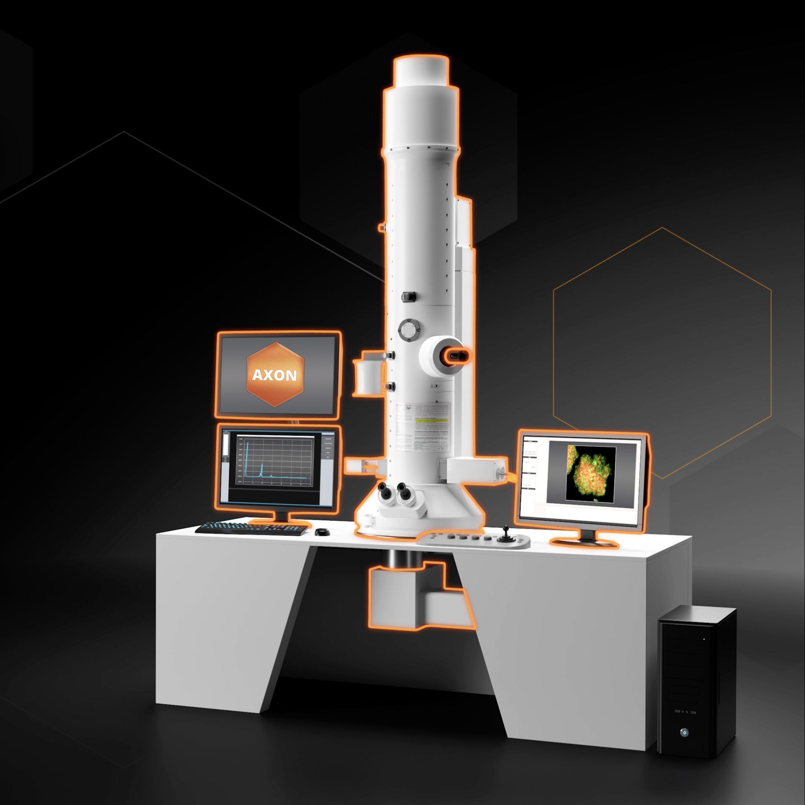
TEM Machine-Vision Software
Protochips' machine-vision software, AXON, redefines the in situ experience by linking the transmission electron microscopy detectors and in situ systems together with a revolutionary software platform. The software collects and saves over 200 metadata that are all indexed. This metadata is linked to the (S)TEM images which can quickly be plotted, exported and published in overlays for videos. It improves data quality, enhances and extends your current microscope capabilities and makes in situ experiments easier for the novice to most advanced users. The AXON platform is a module-based software solution. Easily plug in new modules as they are released, and your system will stay up to date with the latest features.
RDM and FAIR
Publishing research data using Findable, Accessible, Interoperable, and Reusable (FAIR) principles is becoming increasingly important particular when applying for funding.

Click here to find out why