Recently, we traveled to Cambridge, Massachusetts to speak with the microscopy community at Massachusetts Institute of Technology. Along with Dr. Judy Cha of Yale University and Dr. Deborah Kelly of the Virginia Tech Carilion Research Institute, we discussed the wide range of capabilities of in situ TEM holders for various research pursuits.
The day began with a discussion around how monumental in situ TEM holders are, as they transform the TEM from an imaging tool into a dynamic nanoscale world capable of analyzing samples in real-world gas or liquid environments, at high temperature, and in realistic conditions for true operando studies. TEM holders also maintain high resolution within the TEM and are compatible with existing TEM capabilities, such as EDS and EELS.
The discussion then turned to the applications of in situ TEM holders, from observing catalytic reactions in real-time to categorizing nanomaterials in liquid environments. MIT microscopists were intrigued by the capabilities of the Atmosphere gas cell holder, the Poseidon Select liquid cell holder, and the Fusion heating/electrical biasing system holder.
Dr. Judy Cha: Phase Transformations of Nanoscale Systems Using In Situ TEM
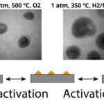
Next, longtime Fusion user Dr. Judy Cha described her experience using the Fusion heating and electrical cell TEM holder in her research. Dr. Cha set out to study how nanoscale phase transformations occur, deviate, and are controlled under thermal or electrical stimuli. However, she required a tool to help her achieve this. By leveraging the capabilities of Fusion, Dr. Cha was able to study IBM’s confined phase change memory (PCM) devices with a metal surfactant layer, as well as metallic glass nanostructures, which are used to test the limits of classic nucleation theories at the nanoscale.
Using the Fusion in situ TEM holder, Dr. Cha observed that the nanorods crystallized at the same rate, no matter their size. As many nanoscale systems rely on phase transformations for switching their functionalities in response to stimuli, the investigation of such phase transformations and subsequent correlation to changes in materials properties are critical.
Dr. Deborah Kelly: Revealing Molecular Adversaries of Human Health
After lunch, we heard from Dr. Deborah Kelly, who uses the Poseidon Select in situ TEM holder in her Cryo-EM lab to analyze biological materials in liquids. Dr. Kelly’s research aims to improve scientific understanding of human health and disease by using in situ cryo-EM to study cells and molecules in detailed native solutions.
Dr. Kelly described how her team uses liquid cell electron tomography and silicon nitride microchips to create environmental chambers for study inside the electron microscope. Using this technique, she observed gold nanorods interacting with glioma stem cells, as well as viewed BRCA1 protein structures in real time. Dr. Kelly said that using in situ TEM holders in the cryo-EM allows her to “look at cancer cells in new ways”.
Fusion Product Demonstration
The final portion of the day took place in one of MIT’s TEM laboratories. Dr. Madeline Dukes led a demonstration of the Fusion heating and electrical biasing cell. Dr. Dukes heated gold within the TEM to demonstrate the capabilities of the Fusion system, as well as the precision of the Clarity software platform that powers it.
Final Thoughts
We enjoyed our day at MIT and loved hearing from colleagues in the microscopy industry about their groundbreaking in situ research. By demonstrating the capabilities of in situ TEM holders, we aim to educate microscopists about the breakthroughs they can make with operando research; as Dr. Kelly said, “it is very different seeing a snapshot than seeing something in motion.”
Learn more about in situ TEM holders and how they can accelerate your research.