It is now possible to visualize at nanometer resolution the infection of a living, biological cell without compromising cell viability using in situ scanning transmission electron microscopy (STEM).
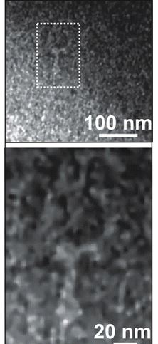
Study: Live Bacterial Liquid Cell Observed Under STEM
To provide contrast while preserving viability, Escherichia coli and P1 bacteriophages were first positively stained with a very low concentration of uranyl acetate in minimal phosphate medium and then imaged with low-dose STEM in a microfluidic liquid flow cell. Under these conditions, it was established that the median lethal dose of electrons required to kill half the tested population was LD50 = 30 e–/nm2, which coincides with the disruption of a wet biological membrane, according to prior reports.
Consistent with the lateral resolution and high-contrast signal-to-noise ratio (SNR) inferred from Monte Carlo simulations, images of the E. coli membrane, flagella, and the bacteriophages were acquired with 5 nm resolution, but the cumulative dose exceeded LD50.
On the other hand, with a cumulative dose below LD50 (and lower SNR), it was still possible to visualize the infection of E. coli by P1, showing the insertion of viral DNA within 3 s, with 5 nm resolution.
The study was published January 26, 2016 in ACS Nano.
Learn more about in situ liquid cell observation with Poseidon Select.


