New Installation Alert at EPFL Switzerland!
April 18, 2024

Studying liquid samples under a transmission electron microscope (TEM) has historically been a challenge for researchers. While TEMs are highly useful for capturing fine details in dry, isolated conditions, the high-vacuum conditions in the microscope prohibit liquid environments. This presents a problem for researchers, since studying samples in their native liquid environments holds countless benefits; imaging samples using in situ liquid cells with electron microscopy can provide unique insights into biological systems, such as cells containing labeled proteins.
It can also provide insight into materials science processes, such as nanoparticle synthesis and electrochemical deposition. Most materials and real-world processes occur in environments containing some moisture, so finding a solution to study these samples with TEM can be critical for researchers.
Specialized liquid cell in situ TEM holders allow researchers to study samples in liquid environments within the TEM. Without these holders, they are unable to visualize materials that naturally exist in liquid environments, such as organic compounds and biological structures.
Let’s take a look at three of the most important benefits of in situ TEM holders for liquid samples:
TEMs require vacuum conditions in order to create and successfully control the precise beam of electrons. While the small wavelength of the accelerated electrons allows the TEM to capture fine detail of the sample, it creates a problem when the researcher is aiming to study samples in real environments.
As water vaporizes under high vacuum, it is necessary to use a vessel that safely seals the liquid layer while remaining transparent to the electron beam. In situ TEM holders for liquid environments accomplish this, enabling the visualization of nanoscale processes in liquid settings.
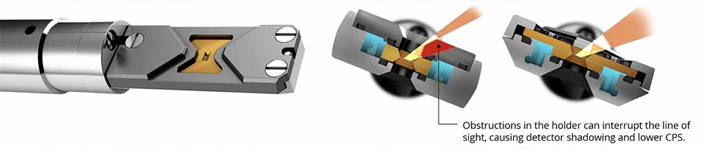
EDS can be incredibly helpful when studying samples under TEM. Since characteristic x-rays emitted from the sample are unique to each element, researchers can determine the composition of samples. Historically, it has been difficult to study samples in liquid environments using energy-dispersive x-ray spectroscopy (EDS); because of the closed-cell design, silicon chips and other materials can block x-rays emitted from samples from reaching the detector.
In situ TEM holders for liquid environments require a design that provides direct line-of-sight from the sample to the detector in order to enable EDS analysis. Recent designs use less material and feature a profile that contains the liquid layer without shadowing the EDS detector and maximizing counts per second (CPS).
A key challenge with studying liquid cells under a TEM is containing the liquid sample. TEMs are extremely expensive, and if liquid escaped from the sample it could potentially destroy the equipment.
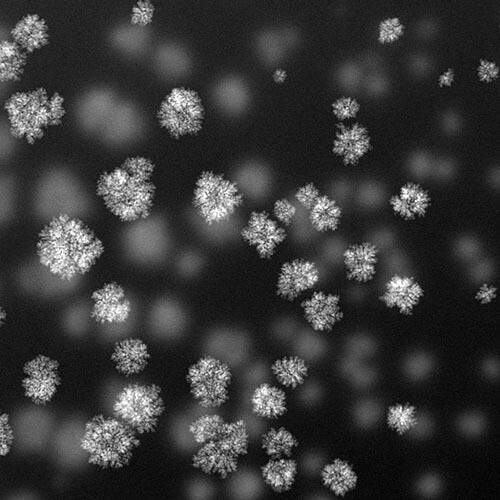
Specialized liquid TEM holders work as sealants, encapsulating liquid dependably within the microscope. These holders use electron-transparent membranes to secure liquids so that observation is not disrupted. This capability is also beneficial for studying live samples, as the holder keeps them encased within an observable window.
In their relatively brief history, in situ TEM holders for liquid environments have revolutionized the research capabilities of the transmission electron microscope. Researchers started out using them to study biological cells, supporting profound observation possibilities, such as whole eukaryotic cells in their native environments.
In the near future, scientists will continue to use liquid in situ TEM holders to branch out into new areas of material science, battery research, corrosion, semi-conductors, cancer research, structural biology, and more.