
Introduction
Energy Dispersive X-ray spectroscopy (EDXS or EDS elemental analysis) refers to the technique of measuring the characteristic x-rays emitted from materials exposed to the high energy electrons used for imaging in SEM and TEM. The ability to conduct EDS Elemental analysis while simultaneously acquiring in situ data and videos has allowed scientists to probe their sample’s nanoscale structural, chemical, and morphological changes in real time. Although EDS is quickly becoming a standard practice of in situ microscopy, there are several factors that can cloud results or impede findings that need to be understood and mitigated.
Counts Per Second (CPS) – The Most Important Factor
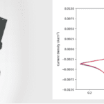
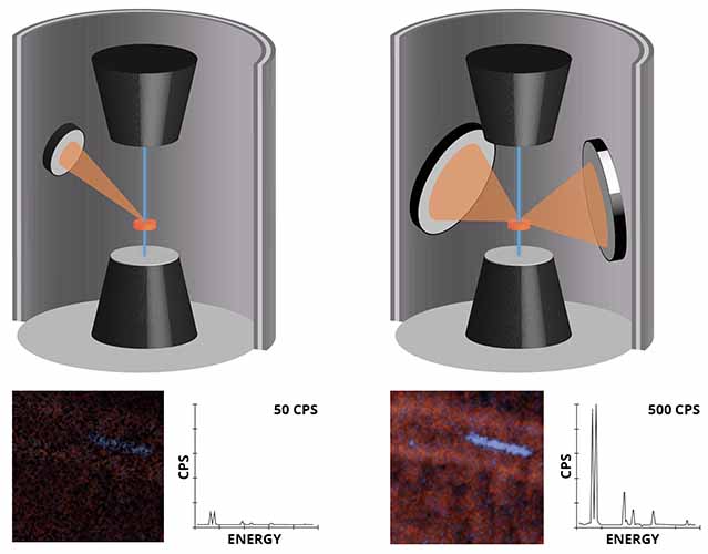
Ultimately, EDS performance and accuracy is dictated by the number of characteristic x-ray counts that are collected. The more x-rays that are collected, the better the EDS results. To maximize CPS, detectors with larger collection angles or with multiple detectors have been developed, and these detectors generally deliver the best EDS results. In addition, new streamlined in situ holders and MEMS devices minimize obstructions to the EDS detectors and improve CPS at any tilt.
Detector Shadowing
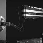
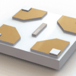
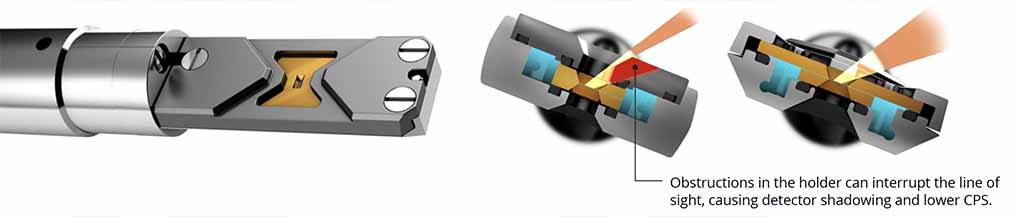
In order for the characteristic x-rays to be collected by the detector, there needs to be direct line of sight from the sample to the detector(s) during collection. As a result, sample geometry, pole piece gap, sample tilt, and holder geometry can all block X-rays from reaching the detector. When the detector is shadowed, the number of characteristic counts per second is reduced or even eliminated, though some non-characteristic counts may be detected from fluorescence. However, you should be able to maximize your sample CPS at any tilt with any detector provided the holder geometry is EDS compatible.
Non-Characteristic Fluorescence
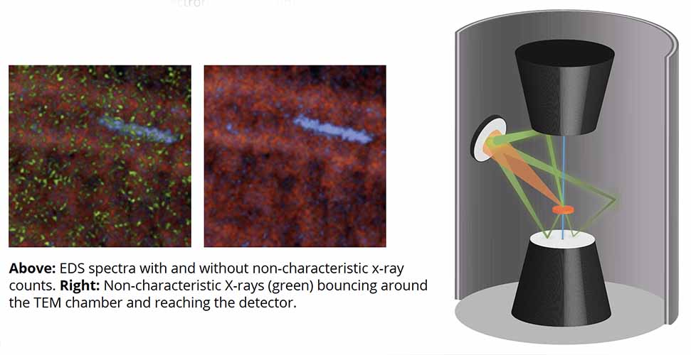
When scattered electrons or x-rays bounce off surfaces in the microscope, they can create secondary x-rays that can be detected by the detectors. As a result, elements like Fe, Cu, Si, and others are often detected even when the detector is shadowed. Therefore, it is important to consider only characteristic x-ray counts when collecting EDS maps.
Conclusion
With many factors affecting EDS elemental analysis results during in situ experiments, holder designs that provide direct line of sight to the detector at any tilt should be utilized. Maximizing the CPS of characteristic x-rays is the safest strategy for optimizing EDS acquisition, so consider the CPS performance of your holder when running experiments.